 |
||||||||||||
| Melanin is the dark pigment present in skin and is produced by melanocytes. It gives our skin its color. Whether dark or light skin, we all have varying amount of melanin. Pigmented lesions occur when melanocytes produce excessive amount of melanin concentrated in one area of skin. Just about every one has a few obvious spots on their skin. Age spots, freckles, moles, melasma and various birthmarks are just few of the commonly known pigmented lesions. High concentration of melanin can be due to various factors. Some types are congenital and present at birth, but most occur with age as a result of sun exposure, pregnancy, hormonal changes and other factors. Most of brown spots are easily ignored but many can detract from your skin’s natural beauty. Fortunately, new state of the art laser, Radiofrequency, chemical peels, microdermabrasion, topical creams and other modalities are available at the Vein Laser Center to lighten or remove these lesions and achieve maximum results. The Medlite Q-Switched YAG laser is one of the most advanced lasers available today for the removal of pigmented lesions. The Medlite delivers the light in very short, high intensity pulses for maximum melanin destruction. The specialized light produced by the MedLite laser is absorbed by the pigmented lesion. The unwanted pigment is then destroyed, thus removing or lightening the lesion. The Medlite Laser is most commonly used to remove brown Age Spots, Freckles, brown birth marks, Nenus of Ota, and Melanocytic nevi. Medlite laser is not used to remove cancerous or suspected of being cancerous. We will evaluate your condition and discuss the appropriate treatment method for you. About 80% of common lesions are removed with one or two treatments. Some deeper lesions will require more treatments. Most common pigmented lesions will not come back but some birthmarks may return months or years later. However, the procedure can be repeated with similar results. |
||||||||||||
Age Spots / Lentigines These are small tan to medium brown macules . They are usually smaller than one cm in diameter. Typically they arise on skin exposed to excessive sun. They increase in size and number as a person ages.THese lesions are treated with Radiofrequency or laser with excellent cosmetic results.
|
||||||||||||
Freckles / Ephelides Small tan macules, 1- 2 mm in diameter, on sun exposed skin. They are seen in people with pale skin and blond or red hair. They initially present in childhood after sun exposure. |
||||||||||||
Café-au-lait macules Tan to pale brown flat patches that often appear at birth or shortly after. They range in size 1 – 20 cm or more. |
||||||||||||
Becker’s Nevi (Hairy nevus) Similar to Café-au-lait patches but they overlie thickened dermis and coarse dark hairs are usually present within the pigmented patch. Mostly it is located over the deltoid or scapular regions. |
||||||||||||
Nevus of Ota & Ito These nevi are common in Asians. Clinically Nenus of Ota appears as a blue-gray patch on the face, usually unilateral, around the eyes, temple, brow, and cheek. (areas supplied by the first and second branches of the trigeminal nerve). Ipsilateral pigmentation of the sclera is also common. Blue—gray color is related to deeper presence of the pigmented cells. Nevus of Ito has the same blue-gray discoloration, but its location is on the shoulder or upper arm. (areas of skin innervated by the posterior supracalvicular and lateral brachial nerves). The pigment in this nevus is deeply placed in the dermis and multiple treatments are needed. |
||||||||||||
Melanocytic Nevi They are acquired or congenital, appear as medium to dark brown macules, occur predominantly on the trunk and extremities. Congenital nevi have been linked with a higher risk of melanoma. |
||||||||||||
Blue Nevi These are blue-black, well circumscribed papules less than one cm in size. It arises spontaneously in children and young adults. They are deeply placed in the dermis. |
||||||||||||
Melasma Is a light to medium brown symmetric face discoloration, most commonly seen on the cheeks, upper lip, nose, forehead and chin. It is associated with pregnancy or oral contraceptives. Usually combination of treatments are required to lighten melasma with high rate of recurrence. Laser, Lightening creams, and Chemical Peels are used at the Vein Laser Center to treat melasma. |
||||||||||||
Post Inflammatory Hyperpigmentation (PIH) Tan to medium brown pigmentation arise in any area of skin that has been traumatized by blunt, sharp or thermal injury. It is common after mosquito bites and scratching. |
||||||||||||
Nevus Spilus They appear on the trunk and extremities as a café-au-lait spots containing darker brown speckles (deep component). |
||||||||||||
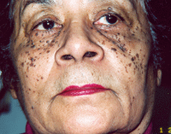
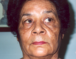
Dermatosis Papulosis Nigra These are small, black marks seen primarily on people of Asian or African descent.
|
||||||||||||
Moles A mole is a flat spot or raised lump on the skin. They vary in size and are usually darker than the skin although they can be pink colored. Moles are more common in lighter skinned people and typically have a hereditary basis. Many birth marks are flat moles. Fortunately, most moles are benign and harmless. Occasionally, however, one can become cancerous. Excessive sun exposure or a history of skin cancer in your family increases your risk of getting skin cancer. You should check all your moles periodically. Look carefully for any of the following:
|
||||||||||||
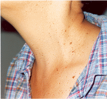
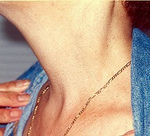
Skin tags These are lesions that do not become cancerous and are often confused with moles. They are usually flesh-colored and are long pieces of skin that stick out. They are not dangerous and can be left alone or, if desired, they can be removed in the office. Since a mole can be mistaken for a skin tag, any change in a skin tag should also be examined by a doctor. |
||||||||||||
Seborrheic Keratoses Seborrheic keratoses are raised non-cancerous growths of the outer layer of skin. They are usually brown, but can vary in color from light tan to black and range in size from a fraction of an inch in diameter to larger than a half-dollar. A main feature of seborrheic keratoses is their waxy, “stuck-on” appearance. A different type of seborrheic keratoses may grow to age spots (solar lentigines). The exact cause of seborrheic keratoses is unknown; however, they seem to run in families. They are not caused by sunlight and can be found on both sun-exposed and non sun-exposed areas. Seborrheic keratoses are more common and numerous with advancing age. Seborrheic keratoses may erupt during pregnancy, following estrogen therapy, or in association with other medical problems. Most often seborrheic keratoses are removed by curettage, electrosurgery, cryosurgery, or laser. |
||||||||||||
Actinic keratoses (solar keratoses) These are considered the earliest stage in the development of skin cancer which is limited to the outermost layer of skin. Since they are caused by the sun, they most commonly occur on body areas such as the face, hands, forearms and the “V” of the neck which are exposed to sunlight. These growths are more common in pale-skinned, fair-haired, light-eyed individuals and are flatter, redder, scalier, and rougher than seborrheic keratoses. |
||||||||||||
Actinic keratoses should be treated. Today, Photodynamic Therapy (PDT) is the treatment of choice for these precancerous lesions. |
||||||||||||


